MEETING & EVENT
HoloAnatomy® and Mixed Reality in the Jagiellonian University
November 2, 2022
HoloAnatomy® Software Suite developed by Case Western Reserve University and Cleveland Clinic is a program based in Mixed Reality (MR) working in HoloLens 2 device. The program was imported as the implementation of the European grant ERASMUS + “MRAME – Mixed Reality supporting Advanced Medical Education –
a new method of teaching medical skills” by its project coordinator, medical engineer Klaudia Proniewska (PhD,Eng). 10 of HoloLens 2 devices were previously purchased by Jagiellonian University by dint of the program “Perfect University- Integrated JU Development”.
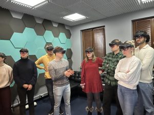
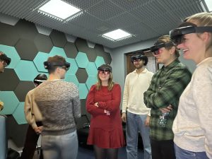
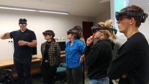
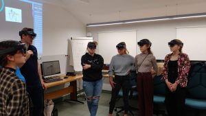
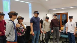
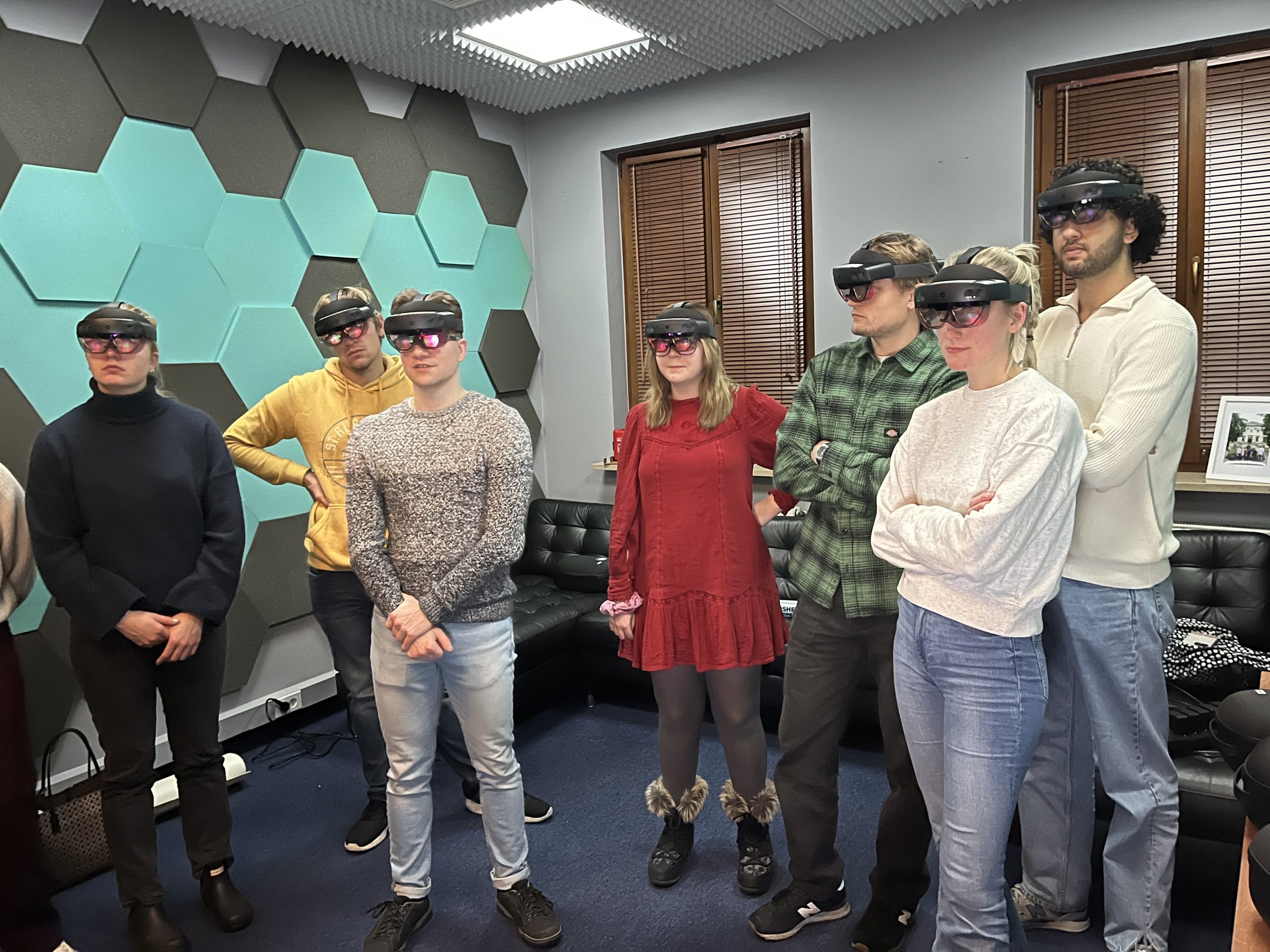
Mixed Reality merges the real world with computer-generated one. Mixed reality is often confused with Virtual Reality, however VR generates only a fully artificial environment whereas MR creates a unique fusion where users can interact with virtual objects in real time. HoloAnatomy® is designed to operate on HoloLens 2 hardware. The headset created by Microsoft stands out with high resolution, voice-control, integration with Microsoft’s softwares and great efficiency. HoloLenses 2 work as an independent, wireless and light-weight personal computer, so users can easily move while wearing them.
HoloAnatomy® Suite takes advantage of these technical benefits and provides students an opportunity to learn anatomy in a completely new way. Heart and soul
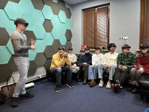
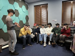
of every HoloAnatomy® lesson is a holographic slideshow. It consists of accurate, three-dimensional models with a controlled number of anatomical structures. There is also a possibility to add other educational materials to the slideshow. What is important, teacher and students see the model in the same place. This enables the tutor to point or magnify any structure from the model he wants to show.
All things considered, HoloAnatomy® not only allows students to better understand anatomy, especially topographical relations between anatomical structures, but also maintains an ability to communicate with the tutor and interact with the model.
JU is the first European university which acquired the HoloAnatomy® Software Suite, joining a centuries-old university tradition with implementation of the latest solutions and technologies in order to refine methods of highly specialized medical staff education. The university has launched pilotage of the HoloAnatomy classes as an addition to the Anatomy course. First year medical students have the opportunity to attend classes in Mixed Reality conducted by experienced anatomists.
It is worth mentioning that students from the School of Medicine in English JU CM (English Division) are also considered in the program and lessons dedicated to them are performed.
Lessons take place in the Mixed Reality Laboratory in the Center for Digital Medicine and Robotics. This space is specially adapted in order to perform classes in a trouble-free way due to stable Wi-Fi connection, matched wall patterns and controlled light intensity. On the other hand, the room remains extremely comfortable and cozy providing students and teachers the best possible experience.
All things considered, implementation of HoloAnatomy® Software Suite allows students to expand their anatomical knowledge gained during classical classes, giving them access to a complete perspective of human topography which is crucial in creating a base for clinical application of anatomy. Moreover, as a part of international cooperation with other medical universities, lessons are planned to be conducted in Radboud University in the Netherlands. The goals are to test a larger number of students and create international questionnaires. As a result, evaluation of educational benefits of HoloAnatomy® Software Suite implementation will be performed.
Our team
HoloAnatomy® lessons are prepared by a creator’s team which is led by medical engineer Klaudia Proniewska PhD and consists of:
-Julianna Dąbrowa, MD Student
-Michał Piotrowski, MD Student
-Antoni Cierniak, MD Student
The classes were conducted by anatomist from Chair of Anatomy CM UJ:
-Professor Jerzy Walocha, MD, PhD, DSc, Head of Department of Anatomy
-Grzegorz Goncerz, MD, PhD
-Bernard Solewski, MD
-Maciej Lis, MD
-Michał Zarzecki, MD
-Michał Goncerz, MSc
Lessons
Topics that have been already completed:
-Joints and ligaments of the upper limb
-Knee joint and lower leg conjunctions
-Joints of pelvis
-Auditory system: external, middle and internal ear anatomy
-Cranial nerves anatomy
-Facial and trigeminal nerves anatomy. Pterygopalatine ganglion
-Spinal cord, meninges and sinuses of dura mater
-Central nerve system. Cerebral circulation
-Thorax: Diaphragm, breast, lymphatic drainage
-Thorax: Mediastinum parts, pretracheal content and vessels
-Thorax: Trachea and Heart (morphology, auscultation, topography)
Topics that are going to be implemented in the near future:
-Muscle of pectoral girdle and arm; axillary fossa, triangular and quadrangular space
-Brachial plexus: nerves, morphology
-Subclavian artery: branches, course. Blood supply to upper limb
-Abdominal wall and muscle
-Stomach and intestines. Abdominal aorta and its branches. Inferior vena cava, portal vein, superior and inferior mesenteric vein
-Celiac plexus and intermesenteric plexus
-Liver, pancreas, spleen; Gallbladder and bile duct
-Kidneys and adrenal glands anatomy. Pelvic floor. Retroperitoneal space content. Bladder. Peritoneum and vessels of pelvis
-Female reproductive system: uterus, fallopian tube, ovary. Vagina: anatomy, blood supply, innervation, lymphatic drainage
-Male reproductive system: testicle, epididymis, ductus deferens, prostate gland, seminal vesicles. Penis: anatomy, blood supply, innervation, lymphatic drainage
-Pelvic muscles; muscular lacuna, femoral canal, obturator canal; muscles of the thigh and lower leg; popliteal fossa
-Lumbar plexus, sacral plexus
-Arteries and veins of lower limb. Inguinal lymph nodes and lymphatic vessels
-Cervical muscles. Cervical plexus and its branches
-Branches and course of cervical vessels. External and internal jugular veins. Common, internal and external carotid arteries. Sympathetic trunk
-Facial muscle, masticatory muscle. Facial artery and vein. Parotid gland
-Retromandibular, temporal, infratemporal, pterygopalatine fossa. Maxillary artery and its branches. Pterygoid plexus. Pterygopalatine, auricular and retromandibular ganglion
-Oral cavity, tongue, retropharyngeal and parapharyngeal space. External nose, nasal cavity. Paranasal sinuses. Larynx
-Visual system: bulb of the eye, eyelid, extraocular muscles and their innervation